Our benefits:
Significantly lower costs
Weekend hours available
Accessible, central location
Top quality radiologists
Same-day results
Appointments and walk-ins
View all the services that Advanced Medical Imaging has to offer across all of our amazing departments

When we hear the word nuclear, most of our minds travel to anxious thoughts of bombs and war, but the word holds a very different meaning in the world of medicine. You may have heard the term “nuclear medicine” thrown around, but what exactly is nuclear medicine?
Nuclear medicine uses radioactive tracers to show the function of different organs within the body. These tracers are most often given to the patient by intravenous injection, although some studies require the swallowing of a radioactive capsule. The camera used in nuclear medicine then detects the gamma rays being emitted from the patient, forming an image on the computer screen. For most studies performed in nuclear medicine, the radiation to the patient is comparable to that of a chest x-ray.
Advanced Medical Imaging (AMI) is the only facility in the Lincoln area to offer DaTscan™, a new test that helps physicians determine if a patient may have a parkinsonian syndrome (PS), the most well-known syndrome of those being Parkinson’s Disease (PD). DaTscan™ is a radiopharmaceutical that is administered prior to a SPECT (single-photon emission computed tomography) scan to help a radiologist see whether or not there is degeneration of dopamine transporters in the brain. Studying this degeneration, along with a patient’s changes in functioning helps physicians to determine if a patient’s symptoms may be related to a PS or rather an essential tremor (ET). Patients interested in finding out if this test could give them the answers they are looking for should ask their neurologist or primary care provider if a DatScan™ at AMI is right for them.
MRI’s of the abdomen are commonly performed to detect issues in the liver, gallbladder, digestive tract, and other organs. It can also be used to evaluate the state of blood vessels and organs prior to a surgery or transplant.
MRI’s of the pelvis are commonly performed to detect issues in the bladder, prostate, reproductive organs, lymph nodes, rectum, anus and pelvic bones.
MRI’s of the breast are many times administered after an initial mammogram to further explore abnormalities. Breast MRI’s are ideal for patients with “dense” breast tissue, because MRI images show more detail allowing radiologists to see more than with a standard x-ray image.
Using an MRI machine to guide a biopsy is very efficient because the clear images help radiologists to accurately sample the abnormality.
MRI is frequently used to scan major joints in the body. Including shoulders, wrists, knees and hips. MRI can locate and identify the cause of pain, swelling, and bleeding in the tissues around joints and bones. The images can see tears and injuries to tendons, ligaments and muscles. MRI can also show arthritis and tumors involving bones and joints.
MRI is frequently used to determine the causes of back pain, leg pain and numbness. The exam can detect a bulging, degenerated or herniated intervertebral disk. MRI can be done to help plan surgeries of the spine. MRI performed after surgery will show whether infection or post-op scarring is present. Patients that have had surgery of the spine may require an injection of contrast material.
MRI of the brain is useful in detecting brain tumors, strokes and certain disorders such as multiple sclerosis. MRI can also detect abnormalities of the eyes or inner ear. Most exams of the brain will require an injection of contrast material to enhance the visibility of certain tissues or blood vessels. A small needle is placed into a vein of the hand or arm for the injection.
MRA provides detailed images of blood vessels with or without the use of contrast material. MRA can detect blocking or narrowing of arteries, and can also detect aneurysms, an enlarged artery. Commonly preformed MRA Exams include brain, carotids (neck) and renal arteries.
Magnetic resonance (MR) enterography is used to diagnose inflammation, bleeding, obstructions and other problems in the small intestine. Patients ingest a barium contrast medium before their scan that highlights certain parts of the digestive tract in the images.
AMI is the only outpatient center in Lincoln equipped with the most advanced MRI that provides highly detailed images, faster. The large bore design is made for patient comfort and is ideal for and larger patients.
Advanced Medical Imaging is the first location in the entire midwest to install a HERO 3T with the latest technology. Scan times are reduced by 50% using Air Recon DL.
The most common of MRI machines is used the most often and has many benefits. There is more signal than the 1T open MRI providing a faster and better image. Less signal than the 3T MRI means less noise and heat than the stronger machine.
The only high-field Open MRI in Lincoln, allows for three times the amount of patient space than cylindrical MRIs. The open design perfect for claustrophobic and bariatric patients.
There are many different studies in nuclear medicine requiring many different preparations by the patient. Most often, your doctor’s office will give you specific directions for your test at the time of scheduling. If you have any questions regarding your test and the preparation required, please feel free to give us a call.
You will generally not have to change out of your clothes for a nuclear medicine procedure, but you will be required for some studies to remove any metal objects. As with any study in radiology, the patient should tell the technologist if there is a chance of pregnancy or if the patient is breastfeeding.
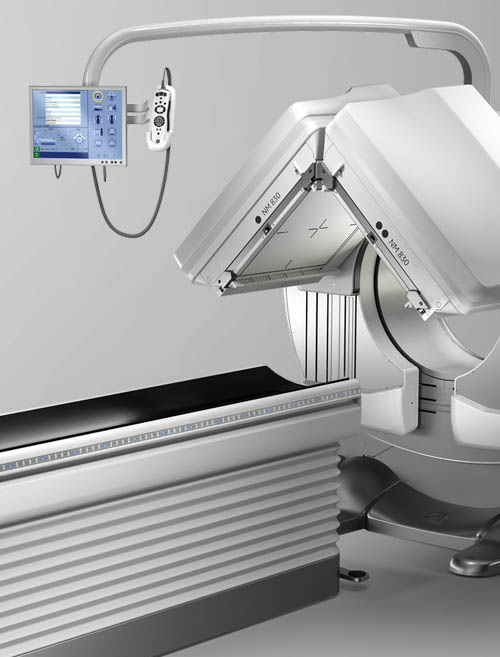
The nuclear medicine gamma camera is a large ring comprised of two cameras that sit 180 degrees of each other. The scanner is open on all sides, and the patient table is positioned in between the two scanners. The cameras are able to move up and down the length of the table, and are also able to rotate around the table. Studies in nuclear medicine vary in time, ranging from several minutes to several hours. Some studies involve scanning at different times during a single day, or some studies are carried out over several days.
A radiologist will determine if there are any abnormalities of the internal organs and bone structures. The radiologist’s interpretation will then be available to your physician 24 hours after your exam or via the patient portal. Your physician’s office will inform you about how to obtain your results.

If you have any further questions about our services, please contact our friendly staff.
Call UsThe short answer is no. Although nuclear medicine procedures do emit a very low dose of radiation, it’s comparable to the amount a person would receive naturally from the environment or from taking a plane trip. It’s important to remember that these exams are only recommended when the benefit far outweighs the risk, so in most cases it is safe to undergo a nuclear medicine procedure.
Anyone can have a nuclear medicine scan if indicated. However, if you are breastfeeding, you may need to stop for a short time after the test. This is due to the small amount of radioactivity in your body that may pass to the breast milk. The nuclear medicine physician will provide you with guidelines. If you are pregnant, we will discuss the benefits and risks of nuclear medicine with you. We use techniques to reduce radiation exposure to the fetus. For instance, we can use smaller amounts of radioactive material for a longer imaging time. In addition, increased hydration and frequent urination can reduce the radiation dose. We will decide whether it is safe before we do the procedure.
You can travel after diagnostic scans. Depending on which scan we perform, there may be a small amount of radiation in your body. This may set off the very sensitive detectors at airports, ferry ports, and train stations. If you have any travel plans, let your technician know so we can give you a card identifying the procedure done and how long sensitive detectors will be able to detect it.

Our Board Certified Radiologists have a wide-variety of subspecialties and are on call 24 hours a day to ensure the best care is always available, when patients need it most.


