Biopsy

When it is not possible to tell from imaging whether a growth is benign or malignant, a biopsy is a highly accurate way to evaluate suspicious masses. Needles are inserted into the abnormal tissue to take a sample for testing. Radiologists use many types of imaging to guide biopsies and get the most accurate samples.  

Biopsies at AMI include:
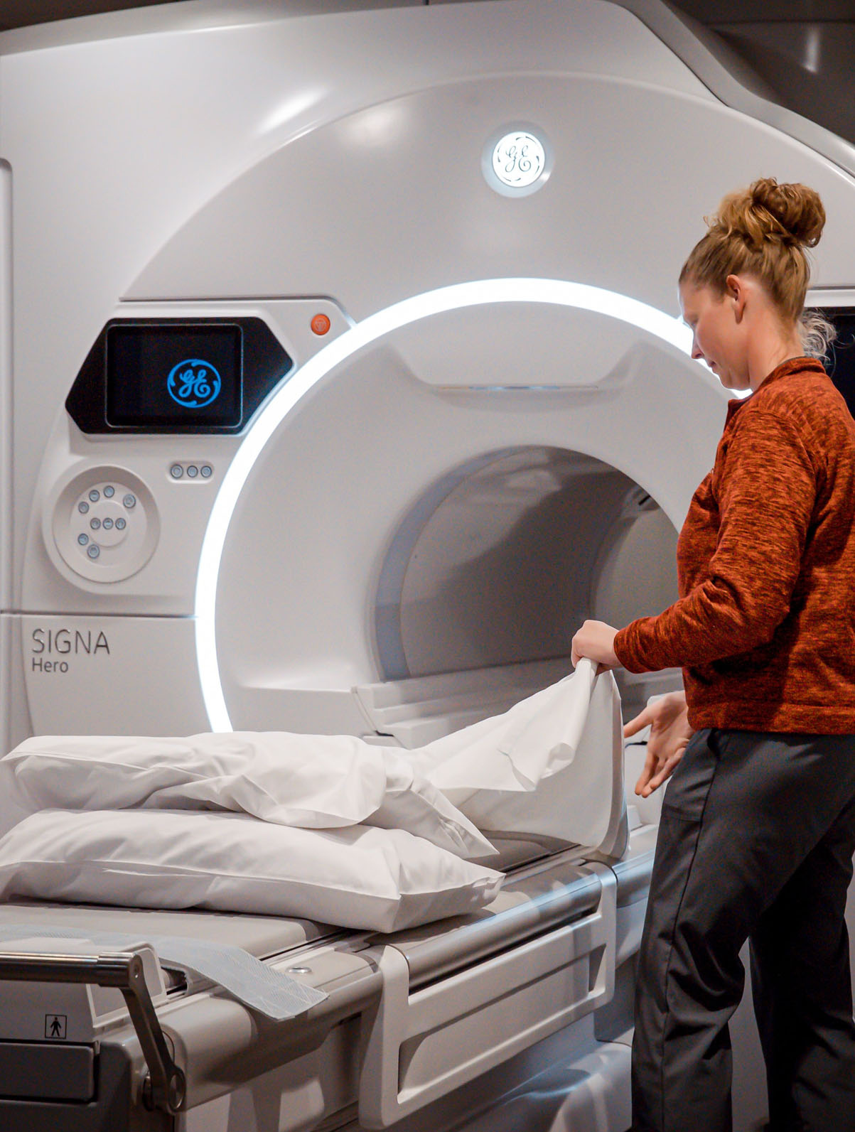
MRI Guided Biopsy
CT Guided Biopsy
Fluoroscopy (X-Ray) Guided Biopsy 

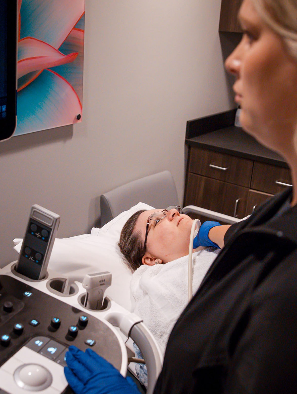
Ultrasound Guided Biopsy

Patients may eat lightly before the biopsy. Patients must not be taking aspirin, aspirin containing products or blood thinners for seven days prior to a needle biopsy.
The radiologist uses lidocaine to numb the area that is to be biopsied. This can sometimes sting for a few seconds but that quickly goes away. After the numbing, patients may feel pressure or pushing but should not feel anything sharp for the remainder of the procedure. An ultrasound is used to locate the lesion and guides a biopsy needle to the mass to be biopsied. About five to seven samples of the area are taken using a spring-loaded device. Each sample will make a “snapping” noise but it should not hurt.

Patients can expect to be at Advanced Medical Imaging for approximately 45 minutes. The actual biopsy will take about 15 to 20 minutes.

Patients will be sent home with a list of simple instructions and are able drive after the procedure. The samples are sent to a laboratory to be evaluated. It takes approximately two working days for the pathologist diagnosis the samples. The results are faxed to the referring physician who will contact the patient regarding the diagnosis.

Stereotactic Breast Biopsy

A stereotactic breast biopsy uses a vacuum assisted needle to obtain breast tissue with mammographic guidance. This tissue is then examined by a pathologist to determine whether it is malignant or benign. A stereotactic biopsy is often performed when a patient has a mammogram showing a suspicious mass, tiny calcifications or an abnormal tissue change.

Patients must not be taking aspirin, aspirin containing products or blood thinners for seven days prior to a needle biopsy.
Clothing from the waist up is removed and patients wear a gown. Patients lie face down on a specially designed table with their breast projecting through a round opening in the table. The table will be raised and the biopsy is performed below the table. A paddle instrument compresses the breast to hold it in place during the procedure.

A radiologist will use lidocaine to numb the area to be biopsied. This can sometimes sting for a few seconds but this quickly goes away. After the numbing, patients may feel pressure or pushing but should not feel anything sharp for the remainder of the procedure. A small nick in the skin is made and the tip of the biopsy needle is advanced to the site of the lesion. X-ray images guide the needle to the right position and then several samples are obtained. A small marker is left at the biopsy site so it can be easily located if the area proves to be malignant. Two mammogram pictures will be taken after the biopsy to serve as baseline images.

Patients can expect to be at Advanced Medical Imaging for 90 minutes. The actual procedure will last for about 30 to 60 minutes.

Patients will be sent home with a list of simple instructions and are able drive after the procedure. The samples are sent to a laboratory to be evaluated. It takes approximately two working days for the pathologist diagnosis the samples. The results are faxed to the referring physician who will contact the patient regarding the diagnosis. If biopsy results are benign, a six-month follow up mammogram will be scheduled.