Our benefits:

Significantly lower costs
Weekend hours available
Accessible, central location
Top quality radiologists
Same-day results
Appointments and walk-ins


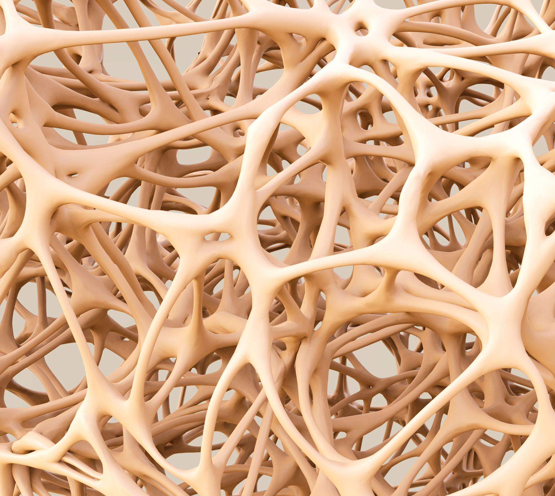
Bone Densitometry is a form of x-ray technology known as dual-energy x-ray absorptiometry (DXA) and is commonly referred to as “DEXA”. DXA is a quick and painless procedure for measuring bone loss. Measurement of the spine and hip are most commonly done. Bone Densitometry is a widely used technique for measuring bone mineral density and diagnosing the presence of osteoporosis.
Osteoporosis can affect anyone, but is most common in post-menopausal women. Bone Densitometry is of limited use in people with spinal deformity or those who have had previous spinal surgery. It is also limited in patients who have had bilateral hip replacements. Osteoarthritis and vertebral compression fractures may also interfere with the accuracy of the test. An order is required for this test.


A DEXA scan gives your physician a baseline for your bone health. Bone loss usually develops gradually and often causes no symptoms, so one measurement only tells part of the story. Comparing a new scan with an earlier one shows whether bone mineral density is holding steady or declining, which is especially useful when a patient is being monitored for osteopenia or osteoporosis.
How often to repeat the test depends on your situation, so your healthcare provider decides the timing. If you've had a bone density scan at another facility, let us know when you schedule so the radiologist has a clear basis for comparison.
A DEXA scan produces measurements, not just pictures. Your report typically includes a T-score, which compares your bone density with that of a healthy young adult and is the standard number used to evaluate osteoporosis. When a prior scan is available, the report can also show how your bone density has changed.
Our bone densitometry service measures bone mineral density, most often at the spine and hip, because those sites are the most useful for assessing bone strength and fracture risk. Talk with the physician who ordered your scan about what your numbers mean and whether any monitoring or treatment is recommended.


MRI’s of the abdomen are commonly performed to detect issues in the liver, gallbladder, digestive tract, and other organs. It can also be used to evaluate the state of blood vessels and organs prior to a surgery or transplant.
MRI’s of the pelvis are commonly performed to detect issues in the bladder, prostate, reproductive organs, lymph nodes, rectum, anus and pelvic bones.
MRI’s of the breast are many times administered after an initial mammogram to further explore abnormalities. Breast MRI’s are ideal for patients with “dense” breast tissue, because MRI images show more detail allowing radiologists to see more than with a standard x-ray image.
Using an MRI machine to guide a biopsy is very efficient because the clear images help radiologists to accurately sample the abnormality.
MRI is frequently used to scan major joints in the body. Including shoulders, wrists, knees and hips. MRI can locate and identify the cause of pain, swelling, and bleeding in the tissues around joints and bones. The images can see tears and injuries to tendons, ligaments and muscles. MRI can also show arthritis and tumors involving bones and joints.
MRI is frequently used to determine the causes of back pain, leg pain and numbness. The exam can detect a bulging, degenerated or herniated intervertebral disk. MRI can be done to help plan surgeries of the spine. MRI performed after surgery will show whether infection or post-op scarring is present. Patients that have had surgery of the spine may require an injection of contrast material.
MRI of the brain is useful in detecting brain tumors, strokes and certain disorders such as multiple sclerosis. MRI can also detect abnormalities of the eyes or inner ear. Most exams of the brain will require an injection of contrast material to enhance the visibility of certain tissues or blood vessels. A small needle is placed into a vein of the hand or arm for the injection.
MRA provides detailed images of blood vessels with or without the use of contrast material. MRA can detect blocking or narrowing of arteries, and can also detect aneurysms, an enlarged artery. Commonly preformed MRA Exams include brain, carotids (neck) and renal arteries.
Magnetic resonance (MR) enterography is used to diagnose inflammation, bleeding, obstructions and other problems in the small intestine. Patients ingest a barium contrast medium before their scan that highlights certain parts of the digestive tract in the images.
AMI is the only outpatient center in Lincoln equipped with the most advanced MRI that provides highly detailed images, faster. The large bore design is made for patient comfort and is ideal for and larger patients.
Advanced Medical Imaging is the first location in the entire midwest to install a HERO 3T with the latest technology. Scan times are reduced by 50% using Air Recon DL.
The most common of MRI machines is used the most often and has many benefits. There is more signal than the 1T open MRI providing a faster and better image. Less signal than the 3T MRI means less noise and heat than the stronger machine.

Wear loose, comfortable clothing. If you avoid clothing with metal zippers, belts or buttons made of metal you will not need to change. Otherwise, you may be asked to change into a gown or scrub pants. Private changing rooms and secured lockers are available. There are no restrictions on what you can eat before the exam.
The exam will last approximately 15-30 minutes. You will lie on your back or side on a padded table to allow for the examination of your spine and hips. Once in position, a detector moves slowly over the area to be imaged. While the exam is in progress, you will be asked to hold as still as possible to ensure a clear image. This process is completely painless.
The results of your bone density exam are interpreted by a radiologist and sent to your doctor and the patient portal. When they receive the results, they will discuss them with you and develop a treatment plan.
If you have any further questions about our services, please contact our friendly staff.
Call UsA DEXA scan measures bone mineral density to help identify bone loss and diagnose osteoporosis. It most commonly measures the spine and hips and gives your physician information about bone strength and fracture risk.
Yes. An order is required for this test. Your healthcare provider will decide whether bone density testing is appropriate based on your medical history, risk factors, or need for follow-up imaging.
Most patients can, but some conditions limit the accuracy of the test, including certain spinal deformities, previous spinal surgery, bilateral hip replacements, osteoarthritis, and vertebral compression fractures. Your provider can determine whether DEXA is the right test for you.

Our Board Certified Radiologists have a wide-variety of subspecialties and are on call 24 hours a day to ensure the best care is always available, when patients need it most.